My podcast is bout oncogene development. Basically I connected all the main points of previous chapters. Starting with the basic definitions of cells and the roles of cell communication and divisions. Then, explaining the two major types of gene mutations which are responsible for tumor suppressing and cell growth leading to cancer development. In addition, I also talked about how these two type of gene’s mutation are dependent on each other and effect the cellular functions causing cancerous tumor.
Chapter 8: Oncogenesis

Oncogenesis, on a lighter note, is the formation of cancer, where the normal cells undergo a transformation, to cancer cells. Cancer is an anomalous cell development that plan to attack different tissues and parts of the human body. Most cells in our body are resting at G1 sub-stage. Notwithstanding, after cells get signals either extracellular signs or intercellular signs, they begin cell division forms. Moving from G1 state to S stage (DNA combination) to G2 stage then M stage. Those strides are firmly managed to keeping any missteps that would prompt terrible results. What’s more, the sorts of transformations include point change, cancellation, insertion, translocation, and enhancement. On the off chance that any of these transformations happens in quality that is dependable in regulation signs for cell division, it prompts cancer. It begins as kind tumor development, non-cancerous, doesn’t attack other tissue, has 1 or 2 changes, and it begins looking changed with time. At that point, it changes to strange cytology, becoming speedier and begins to look not the same as it’s unique. At long last, threatening, cancerous tumor, begins attacking different tissues and looking unique with the first. This reasons a cellular change, which cannot be controlled.
The research problem I addressed is the process by which normal cells are transformed into cancer. I have shown my mastery in my area of research by outlining the step by step process of oncogenesis. Not forgetting that I commenced from the basics of what a cell is and the type of cells, for everyone to comprehend the whole idea.
In conclusion, my readers’ comments describe my work as a great masterpiece. I have come to a conclusion that cancer cells are a great and interesting topic to learn about because there is so much that the public know but so much more that that they do not know on oncogenesis. From my observation, the work is well described, the basics of cells and the comparison in between is well elaborated and precise.
Chapter 7: A New Cancer Treatment That Unlocks the body’s immune system

Melanoma is the most perilous type of skin disease, and these destructive developments create when unrepaired DNA harm to skin cells (largely brought about by ultraviolet radiation or tanning beds) triggers transformations (hereditary defects) that lead the skin cells to duplicate quickly and structure threatening tumors. These tumors start in the color delivering melanocytes in the basal layer of the epidermis.
The article, “Researchers hail new cancer treatment: Unlocking the body’s immune system”, by Don Melvin, CNN, seeks to address a potential breakthrough in skin cancer treatment. The research problem being addressed are cancers that somehow difficult to treat, which include melanoma, advanced lung cancer and cancer that has spread out the body.
It is clear from the article that researchers are doing their job day and night to come up with cancer treatment methods. In fact the author clearly puts it that these potential breakthroughs are not completely employed in dealing with this dangerous disease. I totally agree with the author that the combination of the two drugs – Ipilimumab and Nivolumab – have adverse side effects. He has gone further to clearly saying that the side effects are so adverse that they may offset their benefits in some patients. For this reason, it is evident from the article that this new method cannot replace the existing cancer treatments as much has to be learned on these new drugs. And Nell Barie, a spokeswoman for cancer research further confirms this by putting it clearly that surgery, chemotherapy, and radiotherapy remain vital to cancer treatment. I, however, find it confusing, going by Dr. James Larkin, who claims that the results of these new drugs being a game changer.
Additionally, the author of the article sheds hope by quoting the words of Barrie, where she explains why immunotherapy is the ‘weapon in the arsenal’ in fighting cancer. She explains that cancer, being different from other diseases, is not an invader, instead the body’s cells going rogue. The immune system thus not programmed to target the cancerous cells cannot recognize them as foreign. These drugs hence work in a mechanism that involves switching the immune system back on.
Overall this article is very straightforward. It is a good, well-written article with an important message for both the public and health experts. The piece, when taken as a whole, is relevant and very convincing in theory.
Reference:
http://www.cnn.com/2015/06/01/health/immunotherapy-cancer-treatment-advance/
Chapter 6: Frequently Asked Questions

1. How can gene and DNA mutations be prevented?
Administration of targeted anti-mutagenic agents to our bodies can be used to block DNA and gene mutations during our lifetime. Chlorophyllin is one of the most powerful agents that blocks mutation. Chlorophyllin modifies enzymes that digest carcinogens, it binds with carcinogens so as to speed their excretion, and secure against ionizing radiation.
2. What environmental factors cause cancer?
Unhealthy life choices like smoking, alcohol drinking, poor diets, excess body weight, and no physical activities can cause cancer. Also, exposure to the UV rays and radiation are other environmental causes of cancer.
3. What is the effect of HAMLET on cancer cells?
HAMLET particularly kills cancer cells, and spares cells that are healthy. All tumor cells have been observed to be delicate, while every single healthy cell shows imperviousness to the apoptosis affecting action of HAMLET. HAMLET ties to both insensitive and sensitive cells yet can just enter cancer cells.
4. Are the cancer risks percentages insignificant due to a small number of patients?
These percentages are insignificant not because the sample size of the cancer patients was small, but because not all types of cancers were included. The study justifies itself by acknowledging the fact that they did not include all kinds of cancers and that cancer is caused by a combination of factors.
5. Does inhibiting RAS prior to PI3K inhibit the downward cascade?
Yes, inhibiting RAS prior to PI3K will inhibit the downward cascade. This is because PI3K is upstream of RAS. RAS inhibition induces rapid apoptosis while PI3K inhibition causes tumor regression. For effective antitumor activity, there should be pulsatile inhibition of both RAS and PI3K.
6. Will the utilization of MEK inhibitors always work and will cancer be able to work around this?
Utilization of MEK inhibitors will not always work. This is because some of these inhibitors quickly leave the bloodstream, which makes it hard to keep up enough of the medication in the dissemination to successfully moderate or end tumor development. Others stay in the circulation system too long, which increases the side effects risk.
References
http://www.lifeextension.com/magazine/2009/12/protect-your-genes-from-deadly-mutations/page-01
http://www.cancer.org/cancer/cancercauses/
http://www.acsu.buffalo.edu/~andersh/research/milkcancer.asp
http://www.ncbi.nlm.nih.gov/pmc/articles/PMC4049524/
http://www.cancer.gov/about-cancer/treatment/research/mek
Chapter 5: Novel Allosteric MEK Inhibitor
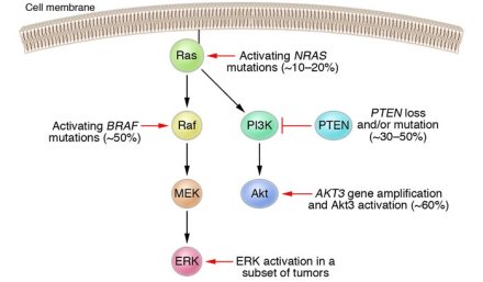
This article relates to cancer cell oncogene as the RAS–RAF–MEK–ERK signalling pathway is hyperactivated in a high rate of tumors, most oftenly inferable from initiating changes of the NF1, RAS and BRAF genes. Research indicates that the utilization of compounds focusing on parts of ERK signaling like RAF or MEK inhibitors has prompted to a substantial change in clinical result in metastatic melanoma and has demonstrated promising clinical activity in extra tumor sorts.
The hypothesis being tested is if the MEK inhibitor, CH5126766 (RO5126766), has the one of a kind property of hindering RAF kinase. Binding of CH5126766 causes MEK to embrace a compliance in which it can’t be phosphorylated by and discharged from RAF. This causes the formation of a steady MEK/RAF complex and restraint of RAF kinase. CH5126766 hinders ERK flagging yield more viably than a standard MEK inhibitor that incites MEK phosphorylation and has strong antitumor movement also. These outcomes recommend that alleviation of RAF input limits pathway hindrance by standard MEK inhibitors.
A scope of cell-surface molecules initiates RAS, a group of GTPases that go about as molecular switches, turning on the downstream RAF protein kinases.The prevailing substrates of RAF kinases are the MAPK/ERK kinases, MEK1 and MEK2. MEK kinases seem to have one and the only primary substrate, ERK. This chain of proteins, from RAS to ERK, conveys signals from cell-surface receptors to the DNA. ERK produces broad changes in quality expression intervened by interpretation figures that control cell cycle movement, separation, protein synthesis, digestion system, cell survival, cell relocation, and attack and senescence.
This study greatly contributes to the knowledge I have for cancer cell oncogene. The part of the ERK signalling
pathway in cancer is seen as more prominent in tumors in which changes in the receptor tyrosine kinases RAS, BRAF, CRAF, MEK1 or MEK2 drive development component autonomous ERK1 and ERK2 enactment and thus improper cell multiplication and survival. New medications that restrain RAF or MEK1 and MEK2 have as of late been affirmed or are presently experiencing late-arrange clinical assessment.
Since CH5126766 adequately restrained ERK phosphorylation in vivo in RAS-mutant xenografts and was a more strong inhibitor of ERK yield and tumor development than PD0325901, does this imply that anticipating actuation of pMEK records for the more prominent viability of this medication?
Reference
Ishii, N., Harada, N., Joseph, E. W., Ohara, K., Miura, T., Sakamoto, H., … & Sakai, T. (2013). Enhanced inhibition of ERK signaling by a novel allosteric MEK inhibitor, CH5126766, that suppresses feedback reactivation of RAF activity. Cancer research, 73(13), 4050-4060.
Chapter 4: PI3K/AKT Pathway

This article is related to cancer cell oncogene on the grounds that the phosphoinositide 3-kinase (PI3K) pathway, a basic sign transduction framework connecting oncogenes and different receptor classes to numerous key cell capacities, is the most normally enacted flagging pathway in human disease. This pathway accordingly introduces a challenge and an opportunity for therapy in cancer.
The hypothesis is if focusing on the phosphoinositide 3-kinase (PI3K) has a restorative worth against human renal cell carcinoma. The outcomes show all chosen human renal cell carcinoma cell lines communicated Akt and showed lifted levels of phosphorylated Akt when developed either in the nonattendance or vicinity of serum, demonstrating a constitutive actuation of the Ser/Thr kinase Akt in human renal cell carcinoma. The outcomes additionally demonstrate that actuation of Akt is tumor particular.
The phosphoinositide 3-kinase (PI3K)/Akt signaling course instigates cell development, cell change, and neovascularization. In vivo bare mice bearing human renal cell carcinoma tumor xenografts were treated with LY294002. Tumor development was measured and tumors were subjected to Western blotch and immunohistochemical investigation. Akt was constitutively enacted in all lines of cells. Constitutive phosphorylation of glycogen synthase kinase-3 (GSK-3) was seen in all cell lines, while forkhead translation element and mammalian focus of rapamycin, albeit communicated, were not constitutively phosphorylated.
This study contributes to my knowledge in cancer cell oncogene. The unusual actuation of the phosphatidylinositol 3-kinase (PI3K)/Akt pathway has been accepted by epidemiological and test ponders as a key stride toward the start and upkeep of human tumors. Some of the intracellular segments of this pathway have been focused as anticancer medication revelation exercises prompting the present panoply of clinical trials of inhibitors of PI3K, Akt and HSP90 in man.
The angiogenic impacts shown with LY294002 in tumors developed in bare mice in this manner also contention the specificity toward Akt of the activities of the PI3K inhibitor. Thrombospondins are communicated in more than 75% of renal cell carcinoma in people. Along these lines, it will be intriguing in future attempts to figure out if the angiogenic impacts we saw with LY294002 in tumors developed in bare mice may be connected to thrombospondin expression.
Reference
Sourbier, C., Lindner, V., Lang, H., Agouni, A., Schordan, E., Danilin, S., & Massfelder, T.(2006). The phosphoinositide 3-kinase/Akt pathway: a new target in human renal cell carcinoma therapy. Cancer research, 66(10), 5130-5142.Reference
Chapter 3 : Cancer Risk Factors

John Hopkins Medicine Research study hopes to change the perception people have on risk factors for cancer. The study’s goal is also to influence cancer studies funding through its results. Out of the 31 cancer patients the study examined, 70.97% cancers were due to bad luck while the other 29.03% was due to either pollution, habits or genetics.
The CNN’s news article header is not appropriate since it claims that the primary cause of most cancers is random gene mutation. This header is different from the John Hopkins Medical Research press release that indicates the study is in explaining the variations in risks of cancer among various types of cancer through the stem cell divisions. The John Hopkins Medical Research is not about the primary cause of many cancers.
The paper’s main focus is on explaining the variation in cancer rates among different tissues. For example, mutations explain why colon cancer is common compared to intestinal cancer. Damaged tissues in our bodies are replenished by the constant division of stem cells in our organs. The more the cells tissues divide, the higher the likelihood of cancer to form in those tissues. An example is the colon tissue; its division is more than the intestinal tissue. Thus, cancer is more prevalent in the colon than in the intestines. Furthermore, I think a more relevant statistic would be to explain the variation in cancer incidence among individuals, for example, why do some individuals get colon cancer while others do not. The new statistic will cover a wide scope of the cancer variations.
Tomasetti & Vogelstein (2015) found that there was a high correlation between the stem cell replication in the tissues and the lifetime cancer risk for those tissues. Brumfield (2015) explains this claim by indicating approximately 65% of random mutation causes cancer risk. Also, the article states that almost a third of the other risks of cancer is due to inherited gene mutations and environmental factors.
The characterization of cancer cells as cells that grow unchecked is intriguing. This implies that the cells keep multiplying until they destroy the host’s organs completely. This may be true in other instances but an important point to note about the cancer cells is that they are not apoptosis. Sometimes random mistakes do occur in the replication of small mutations and DNA. When some genes mutate, they are more likely to promote the growth of cancer. The more the accumulation of these mutations, the higher the risk of unchecked growth cells; this is the cancer hallmark. The cancers that develop have indeed escaped several fail-safe systems in our organs that deal with cell mutations. The fail-safe systems are programmed cell death, DNA repair enzymes and specific immune system cells. If they are not destroyed, they will forever live. Some cancers are slow in growth, and their hosts can normally live for a 2
long time without knowing they have cancer.
Indeed, healthy habits and lifestyle that involves physical exercise, eating healthy foods, and not smoking can help reduce the risk of cancer. This is backed up by the epidemiological data by Dr. Brawley, the chief medical officer for the American Cancer Society. However, cancers due to gene mutations can be best eradicated by early detection when surgery can cure them.
References
‘Bad luck’ can cause cancer, retrieved from file:///E:/Cancer%20%20’Bad%20luck’%20seen%20as%20primary%20cause%20in%20study%20-%20CNN.com.htm
Bad Luck of Random Mutations Plays Predominant Role in Cancer, retrieved from file:///E:/Bad%20Luck%20of%20Random%20Mutations%20Plays%20Predominant%20Role%20in%20Cancer,%20Study%20Shows%20-%2001_01_2015.htm
Chapter 2: cancer cell types
Cancer cell mutation occurs in genes that control:
1- Pro-growth signals / oncogenes cells. Cells think that they had been signaled to permit growing and start the cell division cycle, but they were not signaled by growth hormones.
2- Anti-growth signals / tumor suppressor. Lacking in the expression of tumor suppressor genes.
3- Cell cycle regulation. Over-expression of the proteins that control cell cycle phases.
4- Apoptosis / cell death. DNA damage in the gene(s) that express cell death signals or proteins.

Epithelial tissue
Eighty five percent of human cancer occurs in epithelial tissue, which covers the outside of the body and lines organs and sites within the body. Cancerous cells in epithelial tissue are able to infect other tissues, like connective tissue, resulting in damage and cancer. However, cancer in epithelial cells are called Squamous cells (Carcinoma), which are basically found as the outer skin and lines blood vessels, and columnar and cuboidal cells (Adenocarcinomas) that are found in secretion or active absorption.

Connective tissue
One percent of human cancer is caused by connective tissues that are scattered through an extracellular matrix and functions in holding many tissues and organs together in place. Cancer in connective cells are called Sarcomas. Furthermore, cancerous Fibroblast cells, which secrete fiber proteins, are called Fibro Sarcomas. Cancerous bone forming cells like osteoblasts, osteoclasts and osteocytes, which the skeleton of most vertebrates is made of, are called Osteo Sarcomas. Cancerous lymph cells (5% of cancers) in T-cells and B-cells are called Lymphomas, and cancerous bone marrow cells (3% of cancer), which produce red blood cells, are called Leukemias. Cancerous plasma cells are called Myelomas.

Nervous tissue
Three percent of human cancer presents in nervous tissue cells, which function in the receipt, processing, and transmission of information. This indicates that cancer can start in cells of the brain and spinal cord. Neurons cancer, which is the basic unit of the nervous system, are called Neuroblastomas. In addition, Gliomas and Glioblastomas arise from cancerous Glia, which helps nourish, insulate, modulate, and replenish neuron function.
References:
chapter 1: intro to cell and cancer
cells 
cells is the basic structural, functional, and biological units of all living organisms “building blocks of life”. they are divided into two types: eukaryotic cells which human body, planets, and animals consist of, and prokaryotic cells that bacteria and archaea consist of. all cells share basic features like plasma membrane, and cytoplasm, and genetic materials like DNA and/or RNA that contain genetic information and massages. as a result, they are able to replicate themselves passing down their genetic material to the daughter cells enabling themselves to sustain the cellular functions.

cell communication
communication between cells is essential all multicellular organisms and many unicellular organisms too. cells signal to each other by using chemical signals like hormones and protein. they interpret those receiving signals, process, and respond leading to physical changes, activating down stream signaling by many chemical reactions, cellular regulations, or cell death “apoptosis”.

cell cycle
the ability of living organisms to reproduce themselves and pass down their genetic information through many generations is the best to distinguish between living and non-living organisms. cell division is the processes that all living cells use it to reproduce their own kind, repair, or replacing dead cells. it’s an essential part of the cell cycle, passing down identical genetic material to cellular offspring by using meiosis phase, or division into two daughter cells by using mitotic phase, during growth period. in addition, cell division occur in turn repeatedly through two stages: First, interphase which is divided into sub sub-phases (G1,S “DNA synthesis”, and G2). second, diploid cells “stem cells” go through mitotic phase (prophase, pro-metaphase, metaphase, anaphase, and telophase and cytokinesis) while haploid cells go through meiosis 1 then 2 phases and each phase includes (prophase, metaphase, anaphase, and telophase and cytokinesis).

cancer cell
cancer is an abnormal cell growth which intend to invade other tissues and parts of the human body. most cells in our body is resting at G1 sub-phase. however, after cells receive signals ether extracellular signals or intercellular signals, they start cell division processes. moving from G1 state to S phase (DNA synthesis) to G2 phase then M phase. those steps are tightly regulated to preventing any type of mistakes would lead to bad outcomes. in addition, the types of mutations include: point mutation (nonsense, silent, conservative, and misssense), deletion, insertion, translocation, and amplification. if any of these mutations occurs in gene that is responsible in regulation signals for cell division, it leads to cancer. it starts as benign tumor growth (hyper plastic), non-cancerous, doesn’t invade other tissue, has 1 or 2 mutations, and it starts looking different by the time. then, it transform to abnormal cytology (dysplastic), growing faster, and starts to look different from its original. finally, malignant (neoplastic), cancerous tumor, starts invading other tissue and looking completely different from the original. this causes a cellular transformation (out of control).
Hello world!
This is your very first post. Click the Edit link to modify or delete it, or start a new post. If you like, use this post to tell readers why you started this blog and what you plan to do with it.
Happy blogging!
 abody8y8